Improving Varicose Vein Treatment Accuracy with 3D Imaging
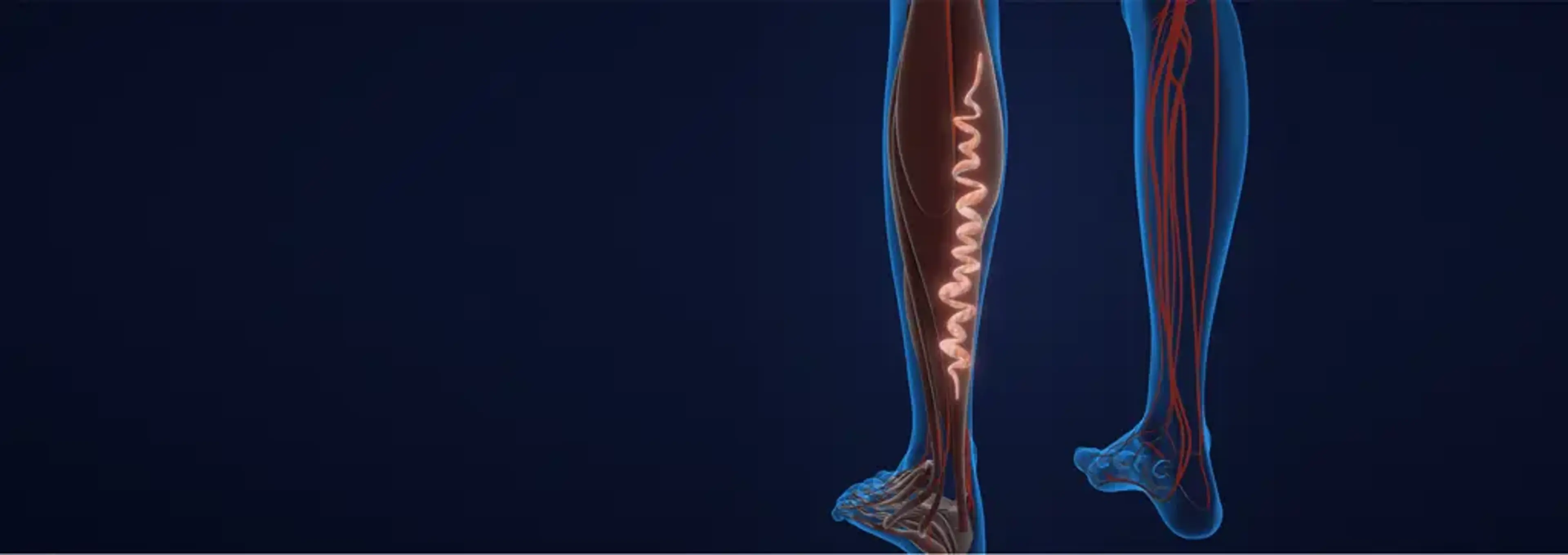
how-3d-imaging-is-helping-to-improve-varicose-vein-treatment-accuracyIf you’ve ever tried to untangle a ball of yarn, you’ll understand one of the challenges vascular surgeons face. Varicose veins don’t run in straight lines. They twist, branch, and hide beneath the skin in ways that are not always obvious from the outside. For years, doctors have relied on duplex ultrasound — an excellent tool — to guide treatment. But ultrasound, like looking at a map in 2D, doesn’t always capture the full picture.
That’s where 3D imaging technology is changing the game. At Charm Vascular Clinic, we see how patients benefit when we move beyond flat images and into three-dimensional vein mapping. It allows us to treat with greater accuracy, minimize complications, and tailor procedures to each patient’s unique vein anatomy.
Why Accuracy Matters in Varicose Vein Treatment
why-accuracy-matters-in-varicose-vein-treatmentTo many people, varicose veins seem like a cosmetic issue — enlarged, twisted veins that appear on the legs. But from a medical perspective, they are often a sign of chronic venous insufficiency (CVI), a condition where valves in the veins no longer close properly. This leads to blood pooling, pressure buildup, and symptoms like:
Heaviness and fatigue in the legs
Pain or throbbing after long periods of standing
Swelling around the ankles
Skin darkening or hardening
In severe cases, venous ulcers
Treating varicose veins is not just about removing what you can see. It’s about addressing the underlying source of reflux. If a surgeon misses the true origin, or if treatment is applied only to visible veins, the problem almost always returns.
Traditional imaging, like ultrasound, provides valuable data but can be limited by:
Operator dependence (results vary depending on skill)
Difficulty visualizing very tortuous veins
Challenges in mapping complex vein junctions
This is why 3D imaging is making such a difference — it allows us to “see the veins in space,” not just in slices.
What Is 3D Imaging in Vascular Medicine?
what-is-3d-imaging-in-vascular-medicine
3D imaging in varicose vein care refers to advanced technologies that reconstruct a three-dimensional map of the venous system. Different systems may be used:
3D Duplex Ultrasound – Builds a volumetric image by stitching together multiple ultrasound slices.
CT Venography (CTV) – Uses computed tomography with contrast dye to visualize both superficial and deep veins.
MR Venography (MRV) – Provides high-resolution 3D vein mapping without radiation, ideal for complex cases.
Augmented Ultrasound Systems – Some clinics now use specialized software that converts real-time ultrasound into 3D visualizations for planning procedures.
At Charm Vascular Clinic, we primarily use 3D-enhanced duplex ultrasound because it’s safe, cost-effective, and patient-friendly. In more advanced or unusual cases — for example, recurrent varicose veins after prior surgeries — we may recommend CT or MR venography for a more detailed map.
How 3D Imaging Improves Treatment Accuracy
how-3d-imaging-improves-treatment-accuracySo, how exactly does 3D imaging translate into better patient outcomes? Let’s break it down.
1. Precise Mapping of Reflux Sources
1.-precise-mapping-of-reflux-sourcesVaricose veins are usually caused by reflux in the great saphenous vein (GSV), small saphenous vein (SSV), or perforator veins. With 3D imaging, we can trace exactly where reflux begins and ends, ensuring treatment targets the true origin rather than just surface branches.
2. Better Planning for Minimally Invasive Procedures
2.-better-planning-for-minimally-invasive-proceduresTechniques like Endovenous Laser Ablation (EVLA) and Radiofrequency Ablation (RFA) rely on inserting a catheter into the vein. 3D maps allow us to plan the entry point, catheter path, and energy delivery with precision, minimizing risks like perforation or incomplete closure.
3. Reduced Risk of Recurrence
3.-reduced-risk-of-recurrenceOne of the main reasons varicose veins come back is incomplete treatment of reflux pathways. By visualizing the full 3D anatomy, surgeons can treat all the problematic segments during one session, reducing recurrence rates.
4. Personalized Treatment Selection
4.-personalized-treatment-selectionNot all veins respond equally well to every technique. For example:
A straight vein may be ideal for EVLA.
A tortuous branch may respond better to foam sclerotherapy.
Large, junctional reflux may need a hybrid approach (ligation + ablation).
3D imaging makes it easier to customize treatment instead of applying a one-size-fits-all solution.
5. Improved Patient Communication
5.-improved-patient-communicationWhen patients can see a 3D map of their own veins, they immediately understand why treatment is needed and what the procedure will address. This builds trust, reduces anxiety, and helps patients commit to follow-up care.
A Real-World Example from Our Clinic
a-real-world-example-from-our-clinicA 47-year-old office worker came to us complaining of leg heaviness and visible varicose veins around her calf. She had previously undergone sclerotherapy at another clinic, but the veins quickly returned.
With traditional ultrasound, her reflux appeared to start from a mid-calf perforator vein. But when we used 3D duplex imaging, we discovered that the true source was reflux in the great saphenous vein higher up in the thigh, which was feeding multiple smaller branches.
By targeting the GSV with endovenous laser ablation, followed by foam for the smaller branches, we treated the root cause. Her symptoms improved within weeks, and she finally experienced long-lasting relief.
Without 3D imaging, her condition may have been misdiagnosed again, leading to yet another recurrence.
3D Imaging and Complex or Recurrent Varicose Veins
3d-imaging-and-complex-or-recurrent-varicose-veinsOne of the most exciting applications of 3D imaging is in complex cases — patients who:
Have already undergone previous procedures
Suffer from large, tortuous vein networks
Have venous ulcers requiring precise planning
Need combination treatments (surgery + ablation + sclerotherapy)
In these patients, a flat 2D view simply doesn’t provide enough clarity. By reconstructing the veins in 3D, we can see connections and hidden branches that would otherwise be missed.
This is especially important in recurrent varicose veins, which often result from untreated tributaries or newly formed reflux pathways. Accurate mapping reduces the likelihood of repeated failures.
Patient Benefits Beyond Accuracy
patient-benefits-beyond-accuracyFrom a patient’s perspective, 3D imaging doesn’t just help the doctor — it improves the entire treatment experience:
Less trial and error – Targeted treatment means fewer procedures.
Shorter recovery times – Because the right technique is chosen from the start.
Lower recurrence risk – Reducing the need for future interventions.
Peace of mind – Patients understand their condition and the plan clearly.
In other words, 3D imaging brings us closer to true precision medicine in vascular surgery.
The Future of 3D Imaging in Vein Care
the-future-of-3d-imaging-in-vein-care
Technology continues to evolve, and we’re only at the beginning of how 3D imaging can transform vascular care. Some of the exciting developments include:
Fusion Imaging – Combining ultrasound with CT/MR data for unparalleled accuracy.
Artificial Intelligence (AI) Analysis – Software that automatically identifies reflux pathways on 3D scans.
Augmented Reality Surgery – Surgeons may soon be able to “see” the 3D veins projected onto the patient’s leg during a procedure, enhancing precision in real time.
At Charm Vascular Clinic, we closely follow these advances because we believe patients deserve the most modern, safest, and most effective care available.
Should Every Patient Get 3D Imaging?
should-every-patient-get-3d-imagingNot necessarily. For straightforward varicose vein cases, a skilled duplex ultrasound may be enough. But 3D imaging is especially valuable for:
Patients with recurrent varicose veins
Those with advanced CVI or venous ulcers
Patients with very tortuous or large veins
People considering hybrid or surgical treatments
In other words, 3D imaging is most helpful when the anatomy is unclear, complex, or high risk.
Final Thoughts
final-thoughtsVaricose veins may look simple on the surface, but inside, they often hide a complicated network of reflux pathways. Traditional ultrasound remains the backbone of diagnosis, but 3D imaging adds an extra layer of accuracy and confidence.
At Charm Vascular Clinic in Seoul, we believe technology should not replace expertise but enhance it. With tools like 3D imaging, combined with Dr. Insoo Park’s surgical experience, we can design treatment plans that are more precise, less invasive, and more durable.
👉 If you’ve struggled with recurrent varicose veins, or if you’ve been told your veins are too complex for simple treatment, consider visiting a specialized center like Charm Vascular Clinic. Advanced imaging may reveal a clearer picture — and a better solution for your long-term vein health.